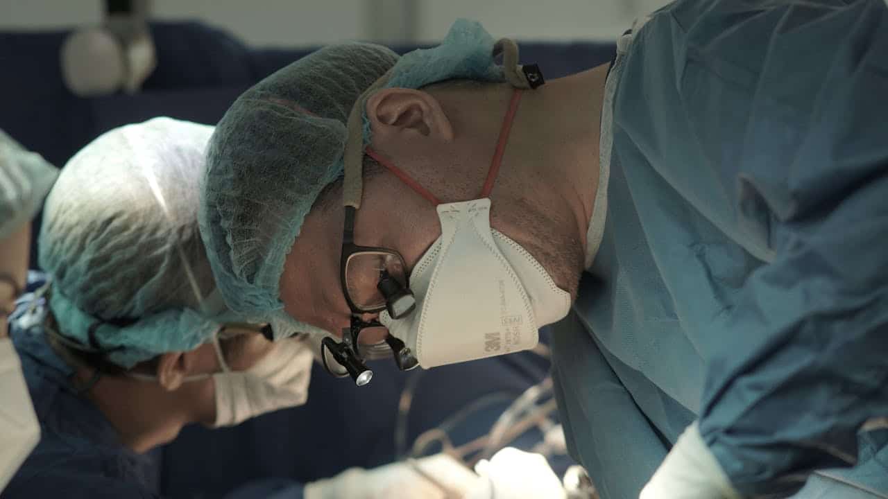
Surgeons at the University of Maryland Medical Center have achieved what seemed impossible: removing a dangerous spinal tumor by going through a patient’s eye socket rather than cutting through the spine. In a world-first operation, neurosurgeon Dr. Mohamed Labib and his team successfully extracted a rare chordoma tumor that had wrapped around 19-year-old Karla Flores’s spine and spinal cord. Only about 300 people receive chordoma diagnoses annually in the United States, making this cancer extremely rare and difficult to treat. Traditional surgical approaches would have required cutting through the back of the neck, risking severe spinal cord damage and paralysis. Instead, doctors pioneered a “transorbital” technique, accessing the tumor through the bottom of the eye socket to create a direct pathway to the cervical spine.
Eye Socket Becomes Surgical Highway to Spine
Dr. Labib developed this innovative approach after recognizing that traditional spinal surgery posed unacceptable risks for Karla’s specific tumor location. Conventional surgery would have required cutting through the back of her neck and potentially damaging the spinal cord to reach the tumor wrapped around her vertebrae.
Working through the eye socket created what Dr. Labib called “a straight shot” to the tumor site. Surgeons accessed the area by making a small incision inside the lower eyelid without disturbing the eye itself. They then removed part of the eye socket floor and cheekbone to create a large enough pathway for surgical instruments.
Dr. Kalpesh Vakharia, the facial plastic surgeon who performed the eye socket reconstruction, carefully cut through the conjunctiva – the transparent membrane protecting the eye – while preserving all visual function. Additional incisions inside the patient’s mouth provided the surgical team with the access they needed.
Endoscopic technology proved essential to the procedure’s success. Surgeons used a thin, lighted tube with a camera to navigate through the newly created pathway and visualize the tumor site. Advanced imaging allowed precise tumor removal while preserving surrounding healthy tissue.

Traditional Surgery Risked Devastating Complications
Approaching Karla’s tumor from the back would have endangered multiple vital structures in her neck area. Surgeons would have needed to work around or potentially damage the spinal cord, which controls movement and sensation throughout the body below the neck level.
Major blood vessels, including the jugular vein and internal carotid artery, run close to the tumor site. Accidentally injuring these vessels during traditional surgery could cause stroke, massive bleeding, or death. The new approach avoided these critical structures entirely.
Nerves controlling swallowing and speech also pass through the area where conventional surgery would have occurred. Damage to these nerves could have left Karla unable to speak clearly or swallow safely, requiring feeding tubes and speech therapy for life.
The eustachian tube, which equalizes pressure between the middle ear and throat, presented another structure that traditional surgery might have disrupted. Damage could have caused permanent hearing problems and chronic ear infections.
Young Woman’s Symptoms Led to Shocking Discovery
Karla first noticed double vision when she turned 18, but spent months trying to identify the cause of her visual problems. Multiple medical consultations failed to provide answers, leaving her feeling frustrated and unheard by healthcare providers who couldn’t explain her symptoms.
An ophthalmologist finally took her concerns seriously and referred her to Dr. Labib for specialized evaluation. Advanced imaging revealed not just one but two separate chordoma tumors – an extraordinarily rare occurrence that medical experts struggle to explain.
One tumor had wrapped around her brain stem, the critical structure controlling breathing, heart rate, and other vital functions. Another tumor encircled her spinal cord in the neck area just below the skull base. Both tumors posed life-threatening risks if left untreated.
Karla described feeling relieved when doctors finally believed her symptoms and took action to help. She had begun to doubt herself after multiple medical encounters failed to identify the serious condition threatening her life.
Multi-Step Surgical Marathon Saves Life
Removing both tumors required three separate operations over several months, each targeting different areas using specialized techniques. Surgeons first addressed the brain stem tumor through traditional skull opening (craniotomy) to remove the largest accessible portion.
A second procedure accessed the remaining brain tumor tissue through the nose using endoscopic techniques. This endonasal approach allowed surgeons to reach areas that the first surgery couldn’t address without causing additional brain damage.
The third and most innovative surgery targeted the spinal tumor through the eye socket pathway. Each procedure was built upon the previous operations to systematically eliminate cancer while preserving Karla’s neurological function as much as possible.
Proton radiation therapy followed surgery to destroy any remaining cancer cells that surgical removal might have missed. Radiation oncologists carefully targeted treatment to affected areas while protecting healthy brain and spinal cord tissue from damage.
Scarless Surgery Preserves Appearance and Function
One remarkable aspect of Karla’s treatment involved the surgical team’s commitment to avoiding external scars. Dr. Vakharia designed the procedure to work entirely through internal incisions that would be invisible after healing.
Titanium plates replaced the removed portions of the eye socket bone, providing structural support while maintaining normal facial appearance. Bone grafts from Karla’s hip reconstructed her cheek area, ensuring proper facial contours after surgery.
The conjunctiva incision inside the lower eyelid heals without visible traces, making it impossible to detect that major surgery occurred. Mouth incisions also heal completely, leaving no external evidence of the complex procedures.
Advanced reconstruction techniques ensured that Karla’s facial appearance remained unchanged despite the extensive bone removal required for tumor access. Her eye socket function returned to near-normal levels, though some minor eye movement limitations persist.

Chordomas Strike Young Adults Without Warning
Chordomas develop from remnants of the notochord, which forms the primitive spine during fetal development. Medical experts don’t understand why these embryonic cells sometimes become cancerous decades later, making prevention impossible with current knowledge.
Most chordomas occur in adults between 20 and 40 years old, affecting people during their most productive life years. The tumors grow slowly but relentlessly, often causing symptoms only after reaching dangerous sizes near vital structures.
Location determines the severity of chordoma complications. Skull base tumors can affect brain function, vision, and hearing. Spinal chordomas may cause pain, weakness, or paralysis depending on their position along the vertebral column.
Having two separate chordomas, as Karla experienced, represents an extremely unusual situation that medical literature rarely documents. Most patients develop single tumors that require complex but more straightforward treatment approaches.
Recovery Brings Hope Despite Challenges
Karla continues recovering more than a year after her surgeries, with no evidence of cancer remaining in her body. Regular monitoring ensures that any tumor recurrence would be detected and treated promptly.
Some lingering effects include limited movement in her left eye due to nerve damage from the brain tumor pressing against her brain stem. While these symptoms may improve over time, they serve as reminders of how close she came to devastating neurological damage.
Spinal fusion surgery stabilized her neck vertebrae after tumor removal, requiring adaptation to some movement limitations. Physical therapy helps maximize her remaining function while protecting the surgical repair sites.
Karla plans to pursue education as a manicurist, moving forward with life goals that seemed impossible during her diagnostic journey. Her positive attitude and determination inspire the medical team that saved her life through innovative treatment.
My Personal RX on Supporting Surgical Recovery
Having witnessed countless patients undergo complex surgeries throughout my medical career, I’m amazed by procedures like Karla’s that seemed impossible just years ago. As someone who believes in the power of both medical innovation and the body’s natural healing capacity, I see her recovery as a testament to what becomes possible when cutting-edge surgery combines with optimal patient care and support.
- Support your body’s healing capacity through optimal nutrition: Focus on anti-inflammatory foods and consider supplements like MindBiotic that support gut health and stress resilience, which are essential for surgical recovery and immune function.
- Prepare your body for surgery with stress reduction techniques: Practice meditation, deep breathing, or gentle yoga to calm your nervous system and optimize your body’s ability to heal from complex procedures.
- Maintain excellent nutrition during recovery: Use recipes from Mindful Meals cookbook that emphasize foods rich in protein, vitamins, and minerals necessary for tissue repair and neurological healing.
- Stay connected with your medical team throughout recovery: Maintain regular communication with all specialists involved in your care, reporting any changes or concerns promptly to ensure optimal healing.
- Prioritize sleep quality to support neurological recovery: Create optimal sleep conditions that allow your brain and nervous system to heal, especially after surgeries involving spinal or brain tissue.
- Engage in appropriate physical activity as cleared by doctors: Follow rehabilitation guidelines carefully while staying as active as safely possible to promote circulation and prevent complications.
- Build a strong support network for emotional healing: Surround yourself with family and friends who can provide practical and emotional support during the challenging recovery period.
- Trust your instincts about your health like Karla did: Advocate for yourself when symptoms persist, seeking additional opinions if necessary until you find healthcare providers who take your concerns seriously.
- Maintain hope while being realistic about recovery timelines: Understand that healing from complex surgeries takes time, and celebrate small improvements while working toward long-term goals.
- Focus on what you can control during recovery: While you can’t control surgical outcomes, you can optimize your nutrition, rest, stress levels, and adherence to medical recommendations to support the best possible healing.
Source:
Graham, F. (2025). Daily briefing: A spinal tumour was removed through a person’s eye socket for the first time. Nature. https://doi.org/10.1038/d41586-025-01441-0


 Subscribe to Ask Dr. Nandi YouTube Channel


