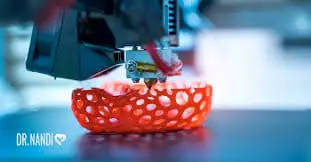
Though 3-D printing seems like a brand new technology, it’s been around for a few decades already. It was in 1981 when Hideo Kodama, a Japanese inventor, used ultraviolet lights to create solid objects. Then in 1984, Charles Hull invented stereolithography. This is a process that allows us to create solid objects using digital data and was the beginning of the 3-D printing era.
Medical Applications of 3-D Printing
Do you remember Y2K? So many things were happening as we lept into the new millennium.
Around this time we also saw the first 3D-printed organ implanted in a human, which was a real breakthrough for scientists from the Wake Forest Institute for Regenerative Medicine. The scientific team printed synthetic scaffolds of a human bladder, then proceeded to coat it with human cells. After that, they were able to successfully implant the new tissue. The incredible thing was there was zero risk of rejection by the immune system since the cells were from the same patient.
During the next decade, 3D-printing continued to be explored within the medical field with favorable results. In a very short time, we saw the fabrication of a miniature kidney, the creation of a prosthetic leg, and the use of human cells to bioprint the first blood vessels. (1)
Stomach Ulcers
Stomach ulcers, also known as gastric ulcers, can result, in part, due to the reduction of the thick protective mucus layer in a person’s stomach. This leaves them vulnerable to the effect of their own stomach acid. According to the CDC (Centers for Disease Controls and Prevention), about 6 million Americans suffer from stomach ulcers every year. (2)
In most cases stomach ulcers are easily treated when detected early, however, in some cases, surgery may be required.
3D Print To Heal Stomach Ulcers
Tao Xu, a bioengineer at Tsinghua University in Beijing, China together with his Ph.D. student Wenxiang Zhao, had an idea to help people suffering stomach ulcers: use 3D printing to re-generate tissue in the stomach lining. (3)
The innovative scientists developed the concept of using “in situ in vivo bioprinting” to treat gastric wounds. The process involves inserting a microrobot into the body, using an endoscope (a tube introduced through body openings). The microrobot then starts printing tissue to repair the damage caused by gastric wall injuries on the stomach.
The microrobot created by the scientific investigators is only 30 millimeters long, but once it is inside the body, it unfolds to 59 millimeters so it can do its bioprinting.
The researchers tested their idea by printing the 2-layer tissue scaffolds on a see-through stomach plastic model. They imitated the anatomical system of the stomach by incorporating human gastric smooth muscle cells and gelatin-alginate hydrogels containing human gastric epithelial cells. Over 10 days, the scientist observed that the printed cells spread steadily, demonstrating the optimal biological function of the cells.
This experiment is not only promising for the bioprinting field but also for the clinical sciences as well. What an exciting time we are living in! What will the future hold?
Sources:


